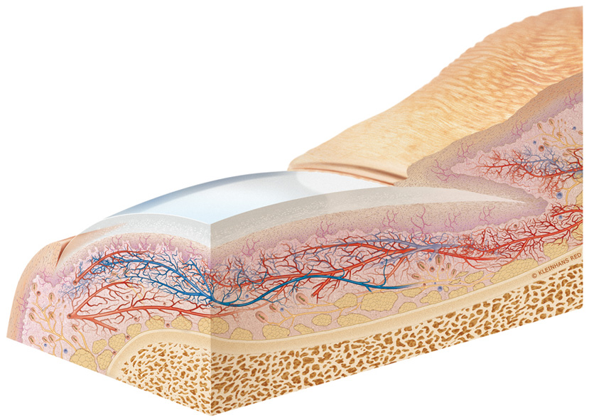
Three-dimensional section of the nail bed

Finger and toe nails develop and rest in the nail bed. They counteract the pressure which is exerted on the tactile elevation. Upon loss of a nail, palpation is limited.
The nail is a modified hornification of the epidermis, a horny plate which is approximately 0.5 mm thick where the nail body and nail root are differentiated. The nail is anchored in the nail bed.
The nail plate is formed by polygonal horny scales. The foremost margin of the nail plate is open, the lateral and behind margins are located in an epithelial fold. The lateral part of the nail plate is located in the groove of the nail bed and the behind one in the nail sinus. Here, the lunula is visible through the nail.
The nail bed is the epithelial tissue which lies underneath the nail. From the matrix, the parent tissue of the nail, the nail grows 0.14 - 0.4 mm each day. The pink color of the nail is due to the underlying capillaries. The color and structure of the nails can be diagnostic tools.
Source: Leonhardt, Helmut. Histologie, Zytologie und Mikroanatomie des Menschen, Band 3, Georg Thieme Verlag 1990, 354-355.
